Get a good grasp on the concepts with the help of free NCERT Solutions at Aasoka. These solutions are prepared in accordance with the latest CBSE syllabus which involves all types of questions along with their solutions. Start practicing with these NCERT Solutions for Class 11 to give a boost to your overall performance. Study, understand, and learn with these solutions written in an easy-to-understand language that can be understood by all students of Class 11.
The chapter “Neural Control and Coordination” of Class 11 Biology includes an explanation of topics like mechanisms of neural coordination, physiology of reflex action, peripheral nervous system, conduction of nerve impulse, nerve impulse transmission, impulse conduction across a synapse, etc.
Question 1:
Briefly describe the structure of the following :
(a) Brain (b) Eye (c) Ear.
Answer:
(a) Structure of brain. The brain is divided into three regions—Forebrain, Midbrain and Hindbrain.
(I) Forebrain. It includes telencephalon and diencephalon.
(1) Telencephalon. It is made up of olfactory lobes and the cerebrum.
(a) Olfactory lobes are concerned with the sense of smell.
(b) Cerebrum is made up of two cerebral hemispheres which are joined by band of nerve fibres called corpus callosum. Each hemisphere is divided into lobes viz. Frontal, Parietal, Temporal and Occipital.
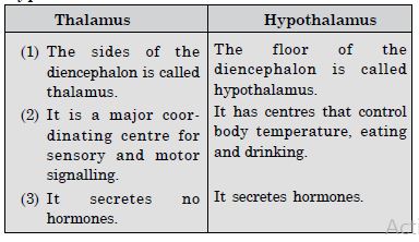
(2) Diencephalon. The roof of diencephalon is called epithalamus. The anterior part of epithalamus is vascular and folded to form the anterior choroid plexus. The epithalamus gives off a body called the pineal body suspended by the pineal stalk. The side walls of the diencephalon are called the thalamus while its floor is called the hypothalamus. The optic nerves coming out of the eyes cross at a point called optic chiasma in front of the hypothalamus. The pituitary gland (hypophysis) is directly attached to the hypothalamus by a stalk called infundibulum.

Structure of Brain (Ventral View)
(II) Midbrain. It exists in the form of two pair of round protrusions called corpora quadrigemina. The anterior pair is called superior colliculi and the posterior pair is called inferior colliculi. Two bundles of fibres lie on the lower surface of midbrain and are called as cerebral peduncles.
(III) Hindbrain. 1. Cerebellum. It consists of two cerebellar hemispheres joined by a central worm-shaped part, the vermis. It controls muscular activities concerned with maintaining body equilibrium.
2. Pons Varoli. It is situated in front of the cerebellum, below the midbrain and above the medulla oblongata. It consists of nerve fibres connecting the two cerebellar hemispheres.
3. Medulla Oblongata. It extends from the pons varoli to the spinal cord. It also possesses a non vascular folded structure called posterior choroid plexus.
(b) Structure of eye. (Fig. 21.8) It is a hollow spherical structure measuring about 2.5 cm in diameter. Its wall is composed of three coats :
(1) Outer fibrous coat made of sclera and cornea.
(a) Sclera. It contain many collagen fibres. It functions to protect and maintain the shape of the eyeball.
(b) Cornea. It is transparent in nature that helps to admit and focus light. It is avascular.
(2) Middle vascular coat made of choroid, ciliary body and iris.
(a) Choroid. It lies adjacent to sclera and is abundant in blood vessels that provide nutrients and oxygen.
(b) Ciliary body. It extends towards the inside of the eye from Choroid. It consists of ciliary muscles and ciliary processes. The ciliary processes secrete aqueous humour. Suspensory ligaments are attached from the ciliary body to the lens and hold the lens in place.
(c) Iris. It is the pigmented region that gives eye its colour. It separates the aqueous humour region into anterior and posterior region. It has an opening in the centre called the pupil. The iris controls the amount of light entering the eye with the help of muscles.

Section of human eye
- Iris provides colour to the eye.
- Lens is an elastic structure made up mainly non-nucleated transparent and elongated cells.
(3) Retina. (Inner nervous coat.)
The retina is the neural and sensory layer of the eye.
- Rods and cones are photoreceptor cells
- Human eye functions as camera.
- Human observe only one object by both the eyes, called binocular vision.
- No rods and cones are present at blind spot.
A small oval yellowish area of the retina lying opposite the centre of the cornea is called
Macula lutea or yellow spot which contains the fovea centralis. The fovea centralis has cone cells only and no rod cells.
(c) Structure of ear. (Fig. 21. 6) Each ear consists of three portions—
1. External Ear. It comprises a pinna and external auditory meatus.
(a) Pinna. The pinna is a projecting elastic cartilage covered with skin. It performs the function of collecting the sound waves.
(b) External Auditory Meatus. It is a tubular passage lined by hairy skin and ceruminous glands. The meatus functions to pass on the sound
waves and ceruminous glands secrete cerumen (ear wax) to prevent the entry of foreign bodies into the ear.
2. Middle Ear. It contains the following parts—
(a) Tympanic Membrane. It separates the tympanic cavity from the external ear. It is thin and semi transparent.
(b) Tympanic cavity. It is the cavity of the middle ear that is connected with the mesopharynx via the eustachian tube that maintains pressure. The tympanic cavity contains a small flexible chain of three small bones called ear ossicles—the malleus (hammer shaped), the incus (anvil shaped) and the stapes (stirrup shaped).
The middle ear is connected to the inner ear through two small openings—
(i) Fenestra ovalis (oval window)
(ii) Fenestra rotunda (round window)
3. Internal Ear. It consists of the membranous labyrinth. The membranous labyrinth or the internal ear consists of the following parts :
(a) Semi-circular canals. There are present three semi circular canals—the anterior, posterior and the lateral semi circular canals which are perpendicular to each other.
(b) Utricle, saccus endoly-mphaticus and saccule. The three semicircular canals are connected to the utricle. The sacculus is joined to the utriculus via a utriculo-saccular duct. From this duct, a long tube called ductus endolymphaticus arises which ends blindly as the saccus endolymphaticus.
(c) Cochlea. It is the main hearing organ which consists of three fluid filled chambers, the upper scala vestibuli, middle scala media and the lower scala tympani. The cochlea contains the organ of Corti which has hair cells that help in hearing.
Question 2:
Compare the following :
- Central neural system and peripheral neural system.
- Resting potential and action potential.
- Choroid and retina.
Answer:
- Differences between central neural system and peripheral neural system.

- Differences between resting potential and action potential.

- Differences between choroid and retina.

Question 3:
Explain the following processes.
- Polarisation of the membrane of a nerve fibre.
- Depolarisation of the membrane of a nerve fibre.
- Conduction of a nerve impulse along a nerve fibre.
- Transmission of a nerve impulse across a chemical synapse.
Answer:
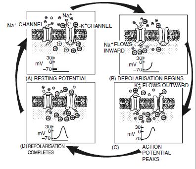
- Polarisation of the membrane of a nerve fibre. The neurons exist in a state of excitability called the polarised state. This is a state of rest when the neuron is not conducting an impulse. At this point, the neurilemma (plasma membrane of a nerve fibre) is more permeable to potassium ions and nearly impermeable to sodium ions. As a result of this, the axoplasm has high concentration of K+ and negatively charged proteins and low concentration of Na+. In contrast, the fluid outside the axon contains low concentration of K+, high concentration of Na+ leading to existence of a concentration gradient.
Such a state of the fibre where there exists an electrical potential difference across the resting plasma membrane is the state of depolarisation and the potential is called the resting membrane potential. - Depolarisation of the membrane of a nerve fibre. The impulse spreads through the length of the nerve fibre in the form of a wave of depolarisation. Depolarisation is a state of reverse polarity. Depolarisation occurs as a result of the opening of Na+ channels whereas K+ channels remain closed. As a result there is an entry of Na+ ions into the neuroplasm which leads to reversed potential difference as inside of the membrane starts turning positive i.e. from –70 mV towards zero. At zero mV, the membrane is said to be depolarized.
- Conduction of a nerve impulse along a nerve fibre. Electrical events in the nerve conduction. The electrical events involve the following changes in the electrical potential of nerve fibre :
- Resting potential state. The nerve cells remain bathed in a fluid called interstitial fluid. In this fluid remain dissolved sodium (Na+) and potassium (K+). During the resting phase, the neurilemma is comparatively 30 times more permeable to potassium ions (K+) than to sodium ions (Na+). As a result Na+ ions are present in high concentration outside the membrane and in low concentration on the inner side. Thus other surface shows (+ve) electric charge and inner surface (+ve) charge due to organic anions. The membrane at this state is said to be in polarised state and the resting potential remains at a value of 70 mm.
- Depolarization and action potential. When a stimulus of any kind is applied to the nerve, it disturbs, the set up. There is marked change in the potential, it is called action potential. The polarity of membrane gets reversed after excitation because the Na+ ions move inward and K+ ions move outward. Thus the membrane is said to be depolarised.
Conduction of nerve impulse
It is termed active phase, during which the inner surface is positively charged and outer surface is negatively charged. - Conduction of impulse. The electrochemical changes are conducted upto synapses as the electric wave of change of potential progresses forward along the fibre.
- Repolarization. In the recovery phase, the axon again restores the original + ve concentration by the outside movement of Na+ ions from the inner side of membrane. The process of change requires something during which nerve cannot be stimulated again. It is of the duration 1/1000 second and called refractory period.
- Transmission of nerve impulse across a chemical synapse Synapse is the close proximity of terminal branch of the axon of one neuron with dendrites of the next neuron in a chain without actual contact. There are two types of synapses i.e. electrical and chemical depending upon the nature of transfer of information across the synapse.
Transmission of nerve impulse across a synapse
Question 4:
Draw well labelled diagrams of Neuron.
Answer:
(a) Neuron.

Structure of neuron
Question 5:
Write short notes on the following—
- Neural Coordination
- Forebrain
- Midbrain
- Hindbrain
- Retina
- Ear Ossicles
- Cochlea
- Organ of Corti
- Synapse.
Answer:
(a) Neural Coordination. The overall functions performed by the nervous system to coordinate and control all the activities of the body is called as Neural Coordination. The brain is the centre of performing physical activities as well as the seat of emotions, will, feelings, intelligence etc. The nerves perform the task of transmitting nerve impulses from the sense organs to the brain and also from the brain to the sense organs.
The Nervous system carries out the function of interaction of two or more organs and complementing the functions of each other.
(b) Forebrain. It consists of cerebrum, thalamus and hypothalamus. Cerebrum is made up of two cerebral hemispheres connected by corpus callosum. Cerebrum is responsible for functions like memory and communication. The cerebrum wraps around a structure called thalamus, which is a major coordinating centre for sensory and motor signalling. Hypothalamus is present at the base of thalamus. It controls body temperature, eating and drinking. It also has neurosecretory cells which secrete hormones.
(c) Midbrain. It consists of cerebral peduncles (bands of nerve fibres) and these lie in the floor of mid brain. These connect the various parts of the fore and hind brain. It consists of two pairs of round elevations, the corpora quadrigemina, act as centres for auditory and visual reflexes.
Functions of mid brain nuclei:
1. Mid brain nuclei control muscle tone.
2. These centres modify motor activities initiated by cerebral cortex.
(d) Hindbrain. It consists of cerebellum dorsally pons varolie and medulla oblongata.
Cerebellum. It lies at the base or posterior part of the brain. The outer cerebellar cortex is made of grey matter. Centrally it has white matter which appears branched. It is not convoluted but is traversed by numerous furrows.
Functions of Cerebellum. (i) It strengthens the impulses to muscles.
(ii) It helps in maintaining tone in muscles.
(iii) It functions in the maintenance of balance.
(iv) It also modulates and moderates voluntary movements initiated by the cerebral cortex.
The pons lies in the front of the cerebellum and above the medulla. Its fibres connect the two halves of the cerebellum and join the medulla with mid brain.
Function of Pons. It co-ordinates the movements of two sides of body.
Medulla oblongata. It lies between the pons and the spinal cord. It is roughly triangular. The medulla contains such vital centres as cardiac, respiratory and arterial pressure.
Functions of Medulla oblongata. (i) It controls the activities of internal organs such as heart, lung and digestive tract, vasomotor centre.
(ii) It also carries nerve tracts between the spinal cord and higher brain centre.
(e) Retina. 1. Retina is the inner sensory coat of eyeball and is formed of following four layers and cells bearing protoplasmic process on inner side.
2. Sensory layer. It lies next to pigmented layer and composed of rods and cones. Rods contain visual pigment rhodopsin and function during dim light. The cones contain pigment iodopsin and function in bright light.
Scattered amongst the rods and cones are supporting cells.
3. Layer of bipolar nerve cells. It is a single layer of neuron. Its dendrites synapse with sensory layer and axon with dendrites of ganglionated layer.
4. Ganglionated Layer. It is the innermost layer of nerve cells. The axons of which converge to form optic nerve.
(f) Ear Ossicles. There are three tiny bones in the middle ear. The middle ear bones called malleus (hammer-shaped), incus (anvil-shaped) and stapes (Stirrup-shaped) form a chain across the middle ear. They extend from the inner surface of ear drum to a similar membrane present on the fenestra ovalis (opening of internal ear).
Function. The ear ossicles transmit the vibrations from tympanic membrane to internal ear after increasing the force.
(g) Cochlea. Cochlea is a spirally coiled duct. It arises from sacculus of vestibule. It is an organ concerned with hearing. If a section of cochlea is cut through, it reveals three tubes as follows :

Structure of Cochlea
Scala vestibuli, scala media and scala tympani. The scala vestibuli is continuous with the vestibuli. The scala tympani is continuous with middle ear at fenestra rotunda. The scala tympani communicates with the scala vestibuli by helico trema. The roof of the scala media is called the Reissner’s membrane while the floor is known as basilar membrane. In this chamber the endolymph is present. On the basilar membrane is present the organ of Corti. It is made up of sensory cells bearing sensory hairs. These hair are embedded in the tectorial membrane arising from basilar membrane. Organ of Corti is the sound perceiving apparatus. Nerve impulse thus set up carry the information to brain.
(h) Organ of Corti. The organ of corti is enclosed at the tip of the cochlean duct in the internal ear. It is formed up of five longitudinal rows of cells (phonoreceptors) held in position by supporting cells. The supporting cells are of two types—long pillar cells and short phalangeal cells. Each auditory or hair cell is a neurosensory cell which has a number of auditory hair on free side and a nerve fibre on other side. The nerve fibres join to form cochlear branch of auditory nerve. The auditory hair are embedded in a gelatinous ribbon like tectorial membrane.
(i) Synapse. (Fig. 21.1) A synapse is a close proximity of end knob of axon of one neuron and dendron or cell body of the next neuron. The former cell is called the Presynaptic cell while the latter is called the Post synaptic cell. The presynaptic cell has numerous mitochondria and synaptic vesicles that contain neurotransmitters. The space between the two cells is called synaptic cleft. There is a release of neurotransmitter such as Acetylcholine from the pre synaptic cell into the cleft by exocytosis. The acetylcholine binds to receptor present over the post synaptic cell. Binding of acetylcholine to its receptor causes depolarisation of the next neuron. To cause inactivation of the process, acetylcholine is inactivated by the enzyme acetylcholinesterase into acetic acid and choline.
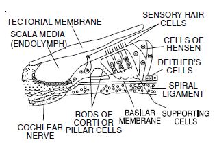
Structure of Organ of Corti
Question 6:
Give a brief account of
- Mechanism of Vision
- Mechanism of Hearing.
Answer:
(a) Mechanism of Vision
1. Formation of Inverted Image on Retina. The rays of light coming from an object pass through cornea, aqueous humor, pupil, lens, vitreous humor and then fall on retina and a small, inverted image of the object is formed.
2. Accommodation. The ability to the eye to focus objects at various distances is accomodation. This is done by changing the convexity of the lens and size of pupil.
3. Photoreception. Light splits rhodopsin pigment into retinene (an aldehyde derivative of Vitamin A) and a protein opsin. This depolarises the rod cells to release a neurotransmitter, transmitting the nerve impulse to different cells and then to optic nerve fibres.
4. Eye to Brain. Nerve impulses are then transmitted from the optic nerve to the brain. The centre for sight is in the occipital lobe of the cerebral hemisphere where an erect image is formed and perceived.
(b) Mechanism of hearing
The phonoreceptors of organ of Corti of cochlea are sensitive to sound waves of frequency ranging from 20 to 20000 cycles/ second or Hertz (Hz) (Maximum sensitive to 1000 cycles per second).
In hearing, different parts of ear perform different functions :
(A) Pinna receives sound waves through its funnel shaped concha.
(B) External auditory canal concentrates the sound waves to increase their pressure to strike and freely vibrate the ear drum.
(C) Middle ear conducts the sound vibrations to fenestra ovalis membrane after increasing their force by about 22 times than at ear drum.
(D) Internal ear
(i) Vibrations of fenestra ovalis cause pressure changes in perilymph of scala vestibuli, vibrations in Reissner’s membrane, pressure changes in endolymph of scala media, vibrations in basilar membrane and pressure changes in perilymph of scala tympani.
(ii) Due to vibrations of basilar membrane, auditory hair phonoreceptors are distorted in the tectorial membrane and are stimulated. The phonoreceptors initiate nerve impulses which are conducted to the auditory area of cerebral cortex of occipital part of cerebral hemisphere. Auditory area interprets the nerve impulses.
(iii) Fenestra rotunda membrane acts as a pressure relief valve in hearing. Its movements are always in opposite direction to that of fenestra ovalis membrane.
(iv) Human ear can differentiate quality as well as quantity of sound waves. Quality of sound waves depends the area in maximum displacement e.g. sound waves of high frequencies (low notes) cause maximum displacement at the tip of basilar membrane while those of low frequencies (high notes) cause maximum displacement at the base of basilar membrane. Quantity of sound waves depends upon the number of phonoreceptors stimulated in a particular area.
Question 7:
Answer briefly :
- How do you perceive the colour of an object ?
- Which part of own body helps us in maintaining equilibrium ?
- How does the eye regulate the amount of light that falls on the retina ?
Answer:
(a) The colour of an object is under the control of the pigment visual violet that is Iodopsin that works in daylight and is sensitive to bright light and colours. There are three different types of pigments under the category of iodopsin :
(i) Erythrolable. It is most sensitive to red light.
(ii) Chlorolable. It is most sensitive to green light.
(iii) Cyanolable. It is sensitive to blue light.
The combination of these three pigments produces all the colours that we can see.
(b) Equilibrium. It is controlled by statoreceptors present in the cristae ampullaes or semicircular ducts and the maculae of vestibule.
Cristae control the dynamic equilibrium (when in motion) and equilibrium during angular acceleration (turning or rotational movements of head). As three semicircular ducts are in three different directions so these can detect the disturbances in position in different directions.
Maculae control the static equilibrium (tilting of head or body at rest) and linear acceleration (rapid forward movements).
(c) The amount of light entering the retina is under the control of two types of muscles present in the iris. These are the circular muscles (sphincters) and radial muscles (dilators). The iris controls the amount of light falling over the retina by the radial muscles contracting in dim light and the circular muscles contracting in bright light.
Question 8:
Explain the following—
- Role of Na+ in the generation of action potential.
- Role of Ca++ in the release of neuro-transmitters at a synapse.
- Mechanism of generation of light induced impulse in the retina.
Answer:
- Role of Na+ in the generation of action potential. The action potential is determined by Na+ ions. The Na+ channels, which are closed in the resting state, open and cause the influx of Na+ ions by diffusion into the inside of axoplasm. The electrical potential of the membrane changes from –70 mV towards zero and then the membrane is said to be depolarised.
- Role of Ca++ ions in the release of neurotransmitters at a synapse. When an impulse arrived at a presynaptic cell, Ca++ ions from the synaptic cleft enter the presynaptic cell. The Ca2+ ions cause the movement of the synaptic vesicles to the surface of the cell and then get fused with the membrane. This is followed by rupturing of the vesicles and release of neurotransmitters of exocytosis into the synaptic cleft.
- Mechanism of generation of light induced impulse in the retina. The photopigments of the retina are photosensitive compounds in the eye that are composed of retinal (an aldehyde of vitamin A) and opsin (a protein). Light induces dissociation of retinal from opsin which leads to a change in membrane permeability. Consequently membrane potential differences are generated producing a signal that generates action potentials in the ganglion cells through the bipolar cells. These action potentials (impulses) are transmitted by the optic nerves to the visual cortex area of the brain where neural impulses are analysed and image is recognised based on earlier memory and experience.
Question 9:
Differentiate between
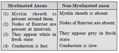
- Myelinated and Non-Myelinated axons
- Dendrites and axons
- Rods and cones
- Thalamus and hypothalamus.
- Cerebrum and cerebellum.
Answer:
- Differences between myelinated axon non-myelinated axon.
- Differences between dendrites and axons.

- Differences between rods and cones.

- Differences between thalamus and hypothalamus.
- Differences between cerebrum and cerebellum.

Question 10:
Answer the following :
- Which part of the ear determines the pitch of a sound ?
- Which part of the human brain is the most developed ?
- Which part of our central neural system acts as a master clock ?
Answer:
(a) Organ of corti of cochlea (b) Cerebrum, (c) Brain.
Question 11:
The region of the vertebrate eye where the optic nerve passes out of the retina, is called the :
- fovea
- iris
- blind spot
- optic chiasma
Answer:
(c) blind spot.
Question 12:
Distinguish between—
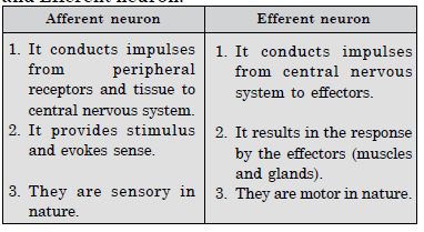
- Afferent neurons and efferent neurons
- Impulse conduction in a myelinated nerve fibre and unmyelinated nerve fibre
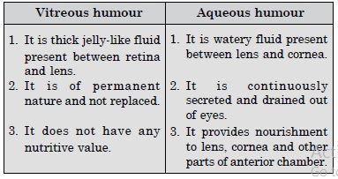
- Aqueous humor and vitreous humor
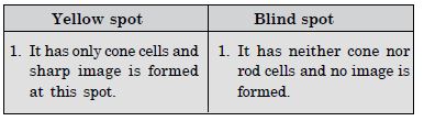
- Blind spot and yellow spot
- Cranial nerves and spinal nerves.
Answer:
- Differences between Afferent neuron and Efferent neuron.
- Conduction of nerve impulse in myelinated and non-myelinated nerve fibre.

- Differences between vitreous humor and aqueous humor.
- Difference between yellow spot and blind spot.
- Difference between cranial nerves and spinal nerves.
