Students of Class 11 can enhance their performance with NCERT Solutions available for free at Aasoka. These solutions aim to provide quality study material designed in accordance with the latest CBSE syllabus. The NCERT Solutions for Class 11 are extremely beneficial to gain knowledge of the concepts related to the subject and chapter. The subject matter experts have written the educational resources in an easy-to-understand language.
In the “Structural Organization in Animals” chapter of Class 11 Biology, students will learn about unicellular and multicellular organisms of the animal kingdom, the functions of various systems (respiratory, digestive, reproductive, circulatory, and nervous) of earthworm, frog, and cockroaches; morphology, the structural organization in animals, etc.
Question 1:
Answer in one word or one line.
- Give the common name of Periplaneta americana.
- How many spermathecae are found in earthworm ?
- What is the position of ovaries in cockroach ?
- How many segments are present in the abdomen of cockroach ?
- Where do you find Malpighian tubules ?
Answer:
- Cockroach.
- 4 pairs.
- Ovaries lie laterally in the 2nd-6th abdominal segments.
- 10
- Malpighian tubules are present at the junction of midgut and hindgut in cockroach.
Question 2:
Answer the following :
- What is the function of nephridia ?
- How many types of nephridia are found in earthworm based on their location ?
Answer:
- Nephridia are excretory structures present in earthworm that regulate the volume and composition of body fluids. A nephridium starts out as a funnel that collects excess fluid from coelomic chamber. The funnel connects with a tubular part of the nephridium which delivers the wastes through a pore to the surface in the body wall or into the digestive tube.
- According to location, there are three types of nephridia :
- Septal Nephridia
- Integumentary Nephridia
- Pharyngeal Nephridia
Question 3:
Draw a labelled diagram of the reproductive organs of an earthworm.
Answer:
Reproductive organs of Earthworm.
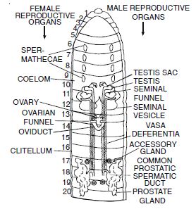
Reproductive system of earthworm
Question 4:
Draw a labelled diagram of alimentary canal of cockroach.
Answer:
Alimentary canal of Cockroach
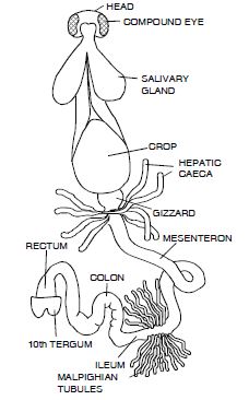
Alimentary canal and associated glands of cockroach
Question 5:
Distinguish between the followings :
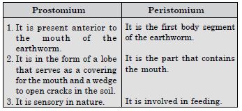
- Prostomium and peristomium
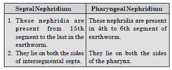
- Septal nephridium and Pharyngeal nephridium.
Answer:
- Differences between pros-tomium and peristomium.
- Differences between septal nephridium and pharyngeal nephridium.
Question 6:
What are the cellular components of blood ?
Answer:
Cellular components of blood.
Blood is a liquid connective tissue made up of fluid matrix plasma and the floating elements i.e. the blood cells and blood platelets.
Cellular components of blood. They are also called formed elements.
(i) Erythrocytes
(ii) Leucocytes
(iii) Blood platelets or thrombocytes.
(i) Erythrocytes. (Red Blood Corpuscles or R.B.C.) The R.B.Cs. are biconcave circular disc without nuclei. The size ranges 7-8 μm. in diameter and 1.5–2 μm thick. The cytoplasm of erythrocytes contains a respiratory pigment, haemoglobin. It is complex protein formed by an Iron-porphyrin ring, called heme and protein globulin. The total number of RBCs per cu mm is 5 million and 4.5 million in adult man and woman respectively. R.B.Cs. are produced in the red bone marrow by a process termed haemopoiesis. If the total count is low, it is termed anaemia and abnormal increase is termed polycythemia.
Erythrocytes have a life span of about 4 months (120 days), afterwards they are destroyed in the liver and spleen. Red blood corpuscles are mainly responsible for transport of oxygen as oxyhae- moglobin.
(ii) Leucocytes (White blood corpuscles or W.B.Cs.). Leucocytes are of different shapes and sizes. They are all nucleated. There are about 6,000–10,000 W.B.Cs. per cu mm of blood of healthy person. In case of infection, the number increases. Leucocytes are actively motile and by virtue of this the shape changes. Leucocytes are produced in the lymph nodes or in the red bone marrow. Length of their active life varies from 3–4 days. Leucocytes are of two types :
(A) Granular Leucocytes
(B) Agranular Leucocytes.
(A) Granular leucocytes or Granulocytes are 9–14 μm in diameter. Their nuclei consist of lobes interconnected by filaments. They are not alike. There are three types of granulocytes according to their staining reaction i.e. Eosinophils, Basophils and Neutrophils.
Eosinophis stain with acidic dye, eosin. They have lobed nucleus. This nucleus stains deep blue. Life span is 14 hours.
Basophils. Cytoplasm have coarse granules and stain with basic dye. Life span is 8–12 days.
Neutrophils. Cytoplasm contains fine dust-like grains. They stain light violet with both acidic and basic dyes. Life span is 10–14 hrs.
(B) Non-granular leucocytes or Agranulocytes. They have no granules in their cytoplasm. They are of two types i.e. Lymphocytes of the diameter 7.5–12 mm with rounded nucleus and Monocytes are large cells having diameter 12–20 mm with bean or oval-shaped nucleus. Life span is short.
WBCs are concerned with formation and storage of antibodies. They attack and engulf the foreign particles.
(iii) Blood platelets. They are very small (2-3 μm) irregularly-shaped structures. The number of platelets varies from 3,00,000 to 5,00,000 per cu mm of blood. They are produced in the bone marrow as they bud off from the cytoplasm of large megakaryocyte. They take part in the clotting of blood.
Question 7:
What are the following and where do you find them in animal body ?
- Chondrocytes
- Axons
- Ciliated epithelium
Answer:
- Chondrocytes. Chondrocytes are the cells of cartilage. These cells are enclosed in small cavities within the matrix secreted by them. A chondrocyte is present in a fluid filled space, the cartilage lacuna.
- Axons. An axon is a single, usually very long process of uniform thickness projecting from a neuron of the nervous tissue.
- Ciliated epithelium. Ciliated epithelium is a type of simple epithelial tissue which is made up of cells bearing delicate hair-like structure called cilia. They function to move particles or mucus in a specific direction over the epithelium. They are mainly present in the inner surface of hollow organs like bronchioles and fallopian tubes.
Question 8:
Distinguish between :
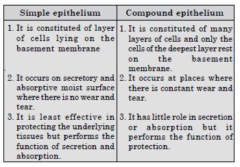
- Simple epithelium and compound epithelium.
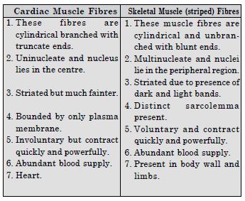
- Cardiac muscle and striated muscle
- Dense regular and dense irregular connective tissues
- Adipose and blood tissue
- Simple gland and compound gland.
Answer:
- Differences between simple epithelium and compound epithelium.
- Differences between cardiac muscle fibres and skeletal muscle fibres.
- Differences between dense regular connective tissue and dense irregular connective tissue
Question 9:
Mark the odd one in each series :
- Areolar tissue ; blood; neuron; tendon
- RBC; WBC; platelets; cartilage
- Exocrine; endocrine; salivary gland; ligament
- Maxilla; mandible; labrum; antennae
- Protonema; mesothorax; metathorax; coxa.
Answer:
- Neuron
- Cartilage
- Ligament
- Antennae
- Protonema.
Question 10:
Match the following
- Compound epithelium
- Compound eye
- Septal nephridia
- Open circulatory system
Answer:
1. (c) 2. (d) 3. (a) 4. (b)
Question 11:
Mention briefly about the circulatory system of cockroach.
Answer:
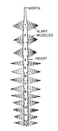
The circulatory system of cockroach is of open type. The visceral organs lie in haemocoel and are immersed in blood called haemolymph. It con-sists of colourless plasma and irregular white corpuscles, the leucocytes. There are no blood vessels except aorta that carries blood from the heart to the haemocoel. The blood is kept circulating by a long, tubular, dorsal, muscular 13-chambered heart. Each chamber of the heart opens into the one in front of it. The opening is guarded by a pair of ventricular valves, which allow only forward flow of blood. At the posterior end of each chamber, except the last, are a pair of holes, the ostia, one on either side and are guarded by auricular valves which allow the blood to pass into the heart from the haemocoel.
Question 12:
Draw a neat labelled diagram of digestive system of frog.
Answer:

Digestive system of frog
Question 13:
Mention the function of the following :
- Ureters in frog
- Malpighian tubules
- Body wall in earthworm.
Answer:
- In male frogs, ureters act as urinogenital ducts i.e. carry urine as well as sperms. In female frogs, ureters only act as urinary ducts.
- Malpighian tubules are responsible for excretion in case of cockroach. These absorb nitrogenous waste product and convert them into uric acid for excretion.
- The body wall in earthworm provides protection, aid locomotion and contains secretory gland cells.